Anatomy lec - 2
Major salivary gland
Parotid gland
•largest one
• Consist of two parts - 1. Main part
2.Accessory part
small detached part, in front of
the main part. superficial to masseter,
aboveparotid duct.
Below zygomatic arch
• Main part :
Site ;
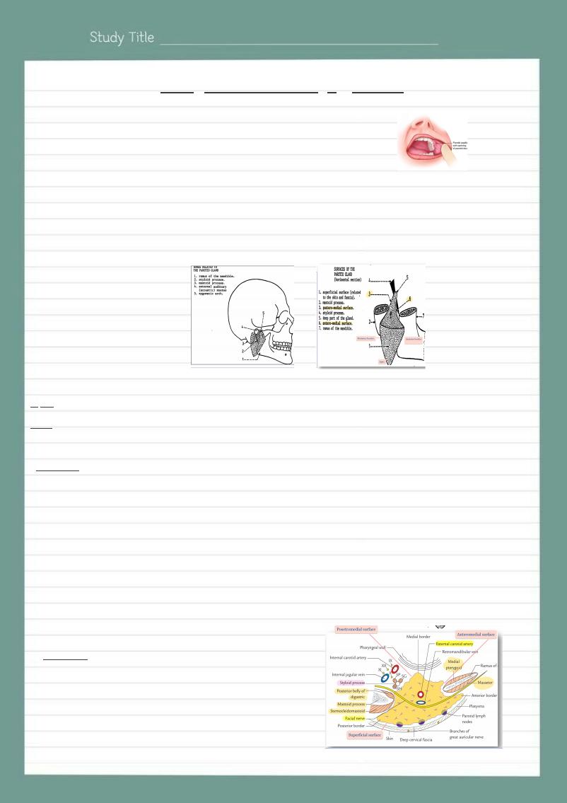
Lodged in gap bounded by :
Above: External auditory meatus.
Anteriorly: Ramus of mandible .
Posteriorly: Mastoid process .
Medially: Styloid process .
Shape ;
3 sided pyramid
Apex (lower end): it is directed downwards.
Base (upper end): it is directed upwards.
3 Surfaces:
1-Lateral surface (superficial).
2-Anterormedial surface (deep).
3-Posteromedial surface (deep).
3 Borders:
1- Anterior border.
2- Posterior border.
3- Medial border
• It gives passage to the two divisions of retromandibular vein.
• related to: cartilaginous part of external auditory meatus and posterior aspect of temporomandibular joint.
• It gives passage to: Superficial temporal vessels and auriculotemporal nerve.
It is related to; Skin, superficial fascia, parotid fascia (investing layer of deep cervical fascia),
superficial parotid lymph nodes.
It is related to; Masseter muscle, Ramus of mandible and Medial Pterygoid muscle.
It gives passage to; Maxillary vessels.
It is related to;
• Mastoid process, sternomastoid and posterior belly of digastric muscles.
• Styloid process and structures attached to it.
• Styloid process separates this surface from carotid sheath and its contents.
It gives passage (entrance) to: External carotid artery and facial nerve.
Parotid duct
• 5 cm
• It emerges from the anterior border of the gland.
• It passes forwards superficial to masseter m
• It pierces the following structures from superficial to deep;
Buccal pad of fat, buccopharyngeal fascia, buccinator muscle and
buccal mucous membrane.
• It opens upon a small papilla in the vestibule of mouth
opposite the upper 2nd molar tooth.