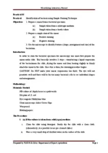


Simple staining in microbiology is a fundamental technique used to visualize bacterial cells under a microscope. This method employs basic dyes such as methylene blue, safranin, and crystal violet to enhance the contrast between the cells and their background. The process involves smear preparation, heat fixing, and staining, allowing for the observation of cell morphology, size, and arrangement. Ideal for microbiology students and professionals, this guide outlines the principles and procedures necessary for effective simple staining. Understanding these techniques is essential for identifying various bacterial groups and their characteristics.
Key Points
- Explains the principle of simple staining using basic dyes to visualize bacterial cells.
- Details the three main steps: smear preparation, heat fixing, and staining.
- Describes common basic stains like methylene blue, safranin, and crystal violet.
- Highlights the importance of heat fixing in adhering specimens to slides.