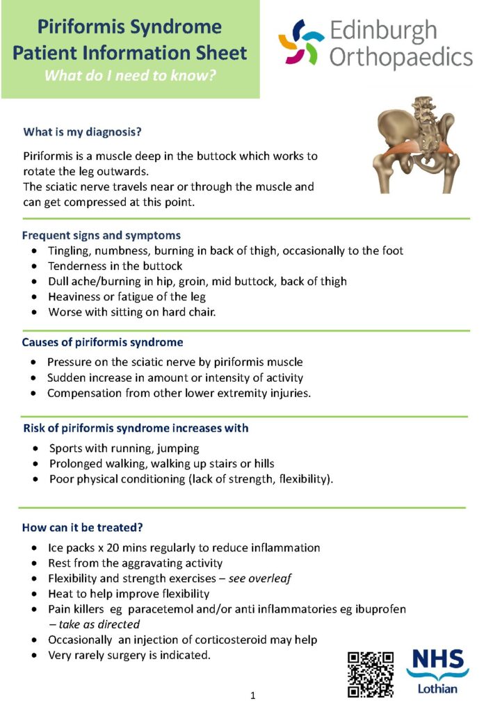


Access an extensive, community-driven archive of muscular system PDFs, sarcomere contraction worksheets, origin/insertion reference tables, and clinical physiology study guides curated to maximize your medical grades and biomechanical understanding. This dedicated resource library tracks the architectural and contractile engines of the human body—ranging from the microscopic precision of actin-myosin cross-bridge cycling and sarcoplasmic reticulum $Ca^{2+}$ release to the macro-level complexity of muscle grouping, torque generation, and postural stabilization. Whether you are troubleshooting the stages of the sliding filament theory, mapping the precise attachment points for limb movement, or preparing for an advanced university physiology or histology test bank, these files give you instant, downloadable clarity.
The Muscular System is the integrated organ network responsible for voluntary movement, postural maintenance, heat production, and the structural integrity of the human frame. Far from a passive series of fibers, the system is a highly complex, electrically-triggered biological engine that converts chemical energy ($ATP$) into mechanical force. Students investigate the system through the lenses of Skeletal Myology (gross muscle anatomy, origins, and insertions), Cellular Physiology (the sarcomeric organization and excitation-contraction coupling), and Tissue Histology (the structural differences between skeletal, cardiac, and smooth muscle types). The field demands extreme precision in identifying muscle groupings, understanding the role of the neuromuscular junction ($NMJ$) in signaling, and mastering the sliding filament theory of contraction. Studying the muscular system builds advanced competencies in biomechanical force modeling, diagnostic clinical evaluation, and multi-system pathology integration—skills foundational to every medical, orthopedic, physical therapy, and athletic training career.
Our collaborative document network hosts student-shared dissection logs, force-production blueprints, and comprehensive board-prep review packages organized across the fundamental branches of myological scholarship:
Systemic Anchoring: Download high-yield muscle origin and insertion tables tracking the primary attachment points for major limb, torso, and facial movements.
Structural Grouping: Access specialized muscle fiber type classification PDFs analyzing the metabolic and fatigue-resistance profiles of Type I (slow-twitch) vs. Type II (fast-twitch) fibers.
Sarcomere Mechanics: Download functional sarcomere contraction diagrams mapping the interaction of actin and myosin, the $Z$-line boundaries, and the sliding filament mechanism.
The Signaling Bridge: Access comprehensive neuromuscular junction physiology guides detailing the release of acetylcholine ($ACh$), the propagation of the muscle action potential, and the role of $T$-tubules.
Tissue Comparison: Download high-yield skeletal vs. smooth muscle histology diagrams highlighting differences in striation patterns, nuclei distribution, and autonomic vs. somatic control.
Clinical Integration: Access dossiers tracking the pathophysiology of conditions like muscular dystrophy, myasthenia gravis, and localized strain injuries.
When analyzing the performance of the muscular apparatus, physiologists rely on standardized biomechanical variables to quantify force and efficiency. The reference matrix below defines the core variables essential for clinical muscular assessment:
| Contractile Variable | Physiological Definition | Clinical / Functional Significance |
| Excitation-Contraction Coupling | The sequence from nerve signal to fiber shortening | Identifying the site of muscle signaling failure |
| Sarcomere Length | The distance between $Z$-lines at rest | Determines the optimal force-production length |
| Motor Unit | A single motor neuron and all fibers it innervates | Governs the precision and strength of contraction |
| Summation | The addition of successive stimuli to increase force | Explains the graduation from twitch to tetanus |
This section addresses the most frequently searched muscular friction points, keyword-targeted contraction prompts, and foundational questions sourced from university medical test banks.
A twitch is the minimal response of a muscle fiber to a single, brief electrical stimulus—a rapid contraction followed by complete relaxation. Tetanus, conversely, occurs when a muscle is stimulated at such a high frequency that there is no time for the fiber to relax between contractions. The successive contractions fuse into one continuous, sustained, and maximal force contraction. This is the mechanism the body uses to maintain prolonged postures or execute high-force movements.
The Sliding Filament Theory dictates that muscle fibers do not physically “shrink” during contraction. Instead, the actin (thin) and myosin (thick) filaments within the sarcomere physically slide past one another. During contraction, myosin heads bind to actin, forming cross-bridges, and perform a “power stroke” using $ATP$ energy to pull the actin filaments toward the center of the sarcomere ($M$-line). As the actin filaments are pulled inward, the $Z$-lines are drawn closer together, and the overall sarcomere length decreases, resulting in the shortening of the entire muscle belly.
Calcium acts as the “on/off” switch for contraction. In a relaxed state, the protein complex troponin/tropomyosin physically blocks the myosin-binding sites on the actin filament. When an action potential reaches the sarcoplasmic reticulum, it triggers the release of stored $Ca^{2+}$. This calcium binds to troponin, causing a structural shift that moves the tropomyosin away from the binding sites. Only then can the myosin heads bind to actin and initiate the contraction cycle. Without calcium release, the muscle remains permanently relaxed.
The nervous system does not contract a whole muscle at once; it recruits “Motor Units.” A motor unit consists of one motor neuron and the group of fibers it activates. For fine, delicate tasks (like moving the eyes), the brain recruits motor units with only a few fibers. For high-force tasks (like lifting a weight), the brain recruits motor units with hundreds or thousands of fibers. Understanding recruitment is essential for understanding how the body scales force and why muscle fatigue occurs as the brain exhausts available motor unit pools.
Yes. Mapping out origin/insertion points, calculating force summation, and debugging complex neuromuscular junction signaling are daily requirements for physiology and medical students. Our global user network frequently uploads complete myology lecture summaries, downloadable sarcomere contraction diagrams, and practice exam answers to help you streamline your study workflow before assessment deadlines.
Every contractile matrix, signaling pathway map, and clinical physiology guide across our database is maintained by a global network of students, researchers, and medical trainees who believe in open, decentralized educational tools. To see how these physiological systems connect with broader anatomical, orthopedic, or pharmacology fields, return to our primary Chesser Resources Browse Directory.
Ready to download premium muscular system study guides or sarcomere contraction diagrams? Join our shared academic network: navigate to your user dashboard, upload 5 of your own physiology lab reports, histology worksheets, or clinical rotation logs, and instantly secure unrestricted access to the high-yield PDFs you need to maximize your medical grades today.